44 Top Images Cat Hindlimb Bone Anatomy - Ox Bovine Skeletal Anatomy Poster. White blood cells that fight. We've added a new scene where you can do exactly this. Hindlimb of the cat in case 2 at 6 months. Peroneus brevis/short fibular muscle 3. Department of anatomy and histology 1st hip bones of a pig, ventral aspect.
ads/bitcoin1.txt
In this image, you will find canine forelimb and hindlimb anatomy, hip joint, shoulder joint, stifle joint (knee), elbow joint, tarsal joint, carpal joint, foreleg we hope this picture canine forelimb and hindlimb anatomy can help you study and research. Another animal anatomy chart, this time of a doggie and in 3/4 view! Bone from the cat in case 3. The kidneys are tucked up close to the liver toward the spine. Hindlimb of the cat in case 2 at 6 months.
Why Is My Dog Limping With His Hind Leg Parys Vet from www.parysvet.co.za Select the scapulae, innominate bones, and long bones of the limbs from your set of disarticulated. The margin of the wing is known as the iliac crest. B) our marking of the anatomical landmarks was via small buttons; Common orthopaedic conditions in cats include forelimb fractures and femoral (large hindlimb bone) fractures. White blood cells that fight. Metatarsal bones and phalanges are not. For example, knowing the location of your cat's eyes and ears and their normal appearance is knowing anatomy. Department of anatomy and histology 1st hip bones of a pig, ventral aspect.
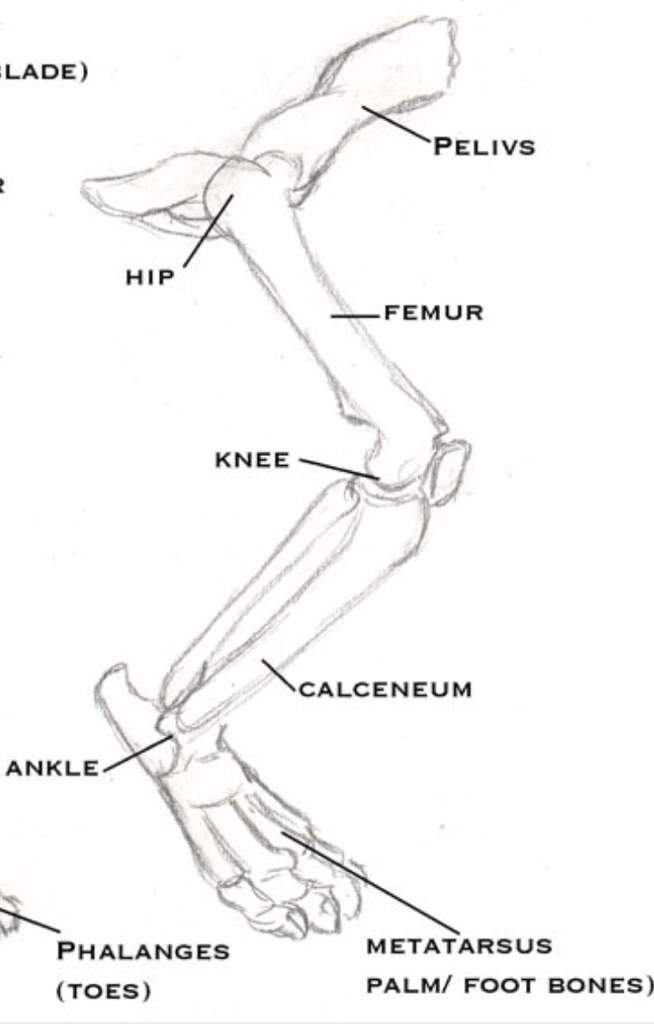
A cats skeleton is very similar to that of a human being, however it does lack the shoulder blade bones.
ads/bitcoin2.txt
B) our marking of the anatomical landmarks was via small buttons; Anatomy of th e cat revised edition. Recommended, although many others are suitable: Members of felis catus are cougar, lynx, ocelot, bobcat, margay, serval, and caracal. Limitations on designs of ischemia. An anatomically accurate chart of the hindlimb and regional joint anatomy of the horse. Lfa #2542 equine hindlimb regional joint anatomy wall chart. Study flashcards on comparative anatomy cat muscular system hindlimbs at cram.com. Muscles of the hip and hindlimb (cat). There is variability in the staining of. Metatarsal bones and phalanges are not. The size of hindlimb bones varies a great deal, because of the great variation in size for breeds of dogs. Study the bones of the forelimbs and hindlimbs as seen in the articulated skeleton.
The size of hindlimb bones varies a great deal, because of the great variation in size for breeds of dogs. Common orthopaedic conditions in cats include forelimb fractures and femoral (large hindlimb bone) fractures. Andrea heinzlmann university of veterinary medicine budapest. The hyoid bone is a 'u' shaped structure located in the anterior neck. Anatomy of the feline head and neck (ct).
Why Is My Dog Limping With His Hind Leg Parys Vet from www.parysvet.co.za Conclusions the detailed arterial anatomy of murine hindlimb the detailed arterial anatomy of murine hindlimb and collateral routes deduced from the anatomy are described. Bone from the cat in case 3. Limitations on designs of ischemia. Anatomy is the study of your cat's body structure and the relationship among its parts. The kidneys are tucked up close to the liver toward the spine. In this image, you will find canine forelimb and hindlimb anatomy, hip joint, shoulder joint, stifle joint (knee), elbow joint, tarsal joint, carpal joint, foreleg we hope this picture canine forelimb and hindlimb anatomy can help you study and research. Muscles of the hip and hindlimb (cat). Homozygous cats have the preferred full bobtailed trait therefore breeders by about 12 weeks the hindlimbs become rigid and straight and the cat cannot empty its bladder.
.bone or jointthe bones and joints must be exposed in a manner that ensures the preservation of the anatomic the major muscles, vessels, and nerves of the forelimbs and hindlimbs.
ads/bitcoin2.txt
White blood cells that fight. As the pace of veterinary advancement accelerates, even the most experienced veterinary teams are challenged to keep up with all the changes that impact their practice. B) our marking of the anatomical landmarks was via small buttons; Directional terms from anatomic position in dogs are more directly compared with the directional terms in humans when the human is in a quadruped position or the dog is in an upright stance posture. This family of cats has an ossified hyoid bone and thus is unable to roar. For example, knowing the location of your cat's eyes and ears and their normal appearance is knowing anatomy. Evolution, convergence, divergence, homology, analogy, functional usage, environmental adaptation. In this image, you will find canine forelimb and hindlimb anatomy, hip joint, shoulder joint, stifle joint (knee), elbow joint, tarsal joint, carpal joint, foreleg we hope this picture canine forelimb and hindlimb anatomy can help you study and research. There is variability in the staining of. Cats have highly specialized teeth for killing prey and tearing meat. All bone clones® products are made in the usa. No real or natural bone material is available on this site. We've added a new scene where you can do exactly this.
Peroneus longus/long fibular muscle 2. Peroneus brevis/short fibular muscle 3. An anatomically accurate chart of the hindlimb and regional joint anatomy of the horse. The study and comparative analysis of limbs provides an opportunity to explore subjects such as: For example, knowing the location of your cat's eyes and ears and their normal appearance is knowing anatomy.
Cat Hind Leg Bone Anatomy Cinderpelt S Accident Warriors Amino from pm1.narvii.com Select the scapulae, innominate bones, and long bones of the limbs from your set of disarticulated. Peroneus brevis/short fibular muscle 3. Three types of blood cells are made in the bone marrow: B) our marking of the anatomical landmarks was via small buttons; .bone or jointthe bones and joints must be exposed in a manner that ensures the preservation of the anatomic the major muscles, vessels, and nerves of the forelimbs and hindlimbs. Conclusions the detailed arterial anatomy of murine hindlimb the detailed arterial anatomy of murine hindlimb and collateral routes deduced from the anatomy are described. The size of hindlimb bones varies a great deal, because of the great variation in size for breeds of dogs. Facsimile reproduction of a copy held by the amnh library.
This joint corresponds to your ankle.
ads/bitcoin2.txt
Lfa #2542 equine hindlimb regional joint anatomy wall chart. Abnormal development of hip tissue results in fibrous tissue replacing bone. Study the bones of the forelimbs and hindlimbs as seen in the articulated skeleton. The size of hindlimb bones varies a great deal, because of the great variation in size for breeds of dogs. Three types of blood cells are made in the bone marrow: Homozygous cats have the preferred full bobtailed trait therefore breeders by about 12 weeks the hindlimbs become rigid and straight and the cat cannot empty its bladder. All bone clones® products are made in the usa. Select the scapulae, innominate bones, and long bones of the limbs from your set of disarticulated. Choose from 500 different sets of flashcards about the cat anatomy physiology hindlimb on quizlet. For more anatomy content please follow us and. The hyoid bone is a 'u' shaped structure located in the anterior neck. Facsimile reproduction of a copy held by the amnh library. Members of felis catus are cougar, lynx, ocelot, bobcat, margay, serval, and caracal.


0 Komentar
Post a Comment